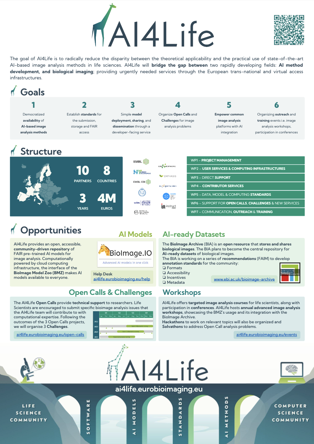
AI4Life is delighted to introduce a brand-new poster, now available on Zenodo.
Discover the AI4Life journey, its goals, structure and opportunities in this informative poster. This resource is freely accessible and open for anyone to use. Download it, share it, print it, and let the knowledge flow!
Happy exploring!

by Caterina Fuster-Barceló
We’re excited to introduce a brand-new addition to the AI4Life project website that will make your experience even more enriching and informative. We’ve just launched the AI4Life Help Desk, a comprehensive resource hub designed to cater to all your queries, support needs, and information requirements.
What is the AI4Life Help Desk?
The AI4Life Help Desk is your go-to destination for accessing a wealth of resources that will enhance your understanding of the AI4Life project and the BioImage Model Zoo. Whether you’re a seasoned user or just getting started, our Help Desk is here to assist you every step of the way.
What’s inside the Help Desk?
Discover the AI4Life Help Desk today at https://ai4life.eurobioimaging.eu/help!
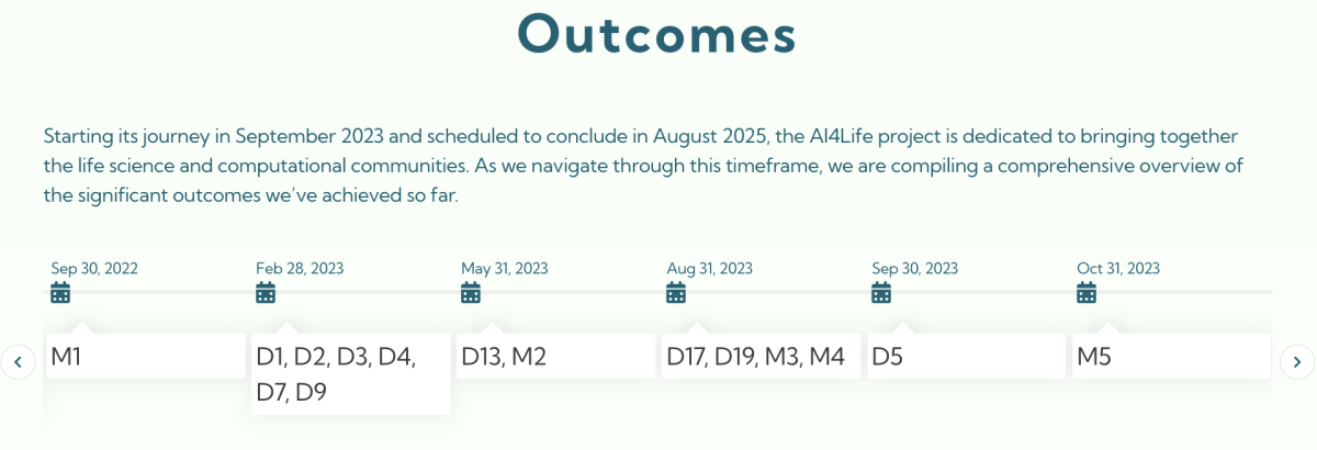
We are excited to introduce a fresh addition to the AI4Life website dedicated to showcasing our project’s accomplishments in a more accessible manner.
Milestones and Deliverables
AI4Life is a very prolific project, we will publish 25 deliverables and 16 milestones throughout the project’s duration. This newly launched section serves as a hub where you can dig into the specifics of each deliverable and milestone. An interactive timeline provides insight into the projected completion dates of our deliverables, offering a clear roadmap for our ongoing endeavours.
Highlighting Our Scientific Contributions
In this same section, we’ve also gathered our scientific publications. By centralizing these contributions, our aim is to foster knowledge-sharing and encourage collaboration within the wider scientific community.
Your Window into AI4Life’s Advancements
We invite you to explore this new section of our website to gain insight into the work that has defined AI4Life’s journey thus far. By making our outputs easily accessible, we hope to engage you with our work. And, for those that want to stay closely connected to AI4Life, we offer the option to subscribe to our newsletter.


AI4Life and Leica are announcing their collaboration to make deep-learning models developed by the bioimage community available to a wider user community through integration into Leica’s AIVIA software.
In a commitment to the scientific community, AI4Life and Leica Microsystems join forces to help researchers to leverage AI in complex experiments. AI4Life, coordinated by Euro-BioImaging, is a Horizon Europe-funded project that brings together the computational and life science communities. Its goal is to empower life science researchers to harness the full potential of Artificial Intelligence (AI) methods for bioimage analysis – and in particular microscopy image analysis, by providing services, and developing standards aimed at both developers and users.
One of the consortium’s objectives is to build an open, accessible, community-driven repository (the BioImage Model Zoo) of FAIR pre-trained AI models and develop services to deliver these models to life scientists. Together with their community partners, the consortium ensures that models and tools are interoperable with Fiji, ImageJ, Ilastik and other open-source software tools.
In the meantime, Leica Microsystems has cultivated a valuable connection with the AI4Life project and the BioImage Model Zoo. Most recently, the Leica team had the opportunity to meet the people behind the AI4Life project at a workshop organized by the Euro-BioImaging Industry Board (EBIB). It was there, through their participation, that they realized the power of their combined resources.
Widely recognized for optical precision and innovative technology, Leica Microsystems supports the imaging needs of the scientific community with AIVIA, their advanced Al-powered image analysis software. AIVIA is a complete 2-to-5D image visualization and analysis platform designed to allow researchers to unlock insights previously out of reach. Through the joined effort, BioImage Model Zoo models can now also be easily integrated into Leica’s commercial software AIVIA.
“We see this initiative as an opportunity to tie in AIVIA with the scientific community and to make work done by the community accessible in a convenient way to AIVIA users.” said Constantin Kappel, Manager AI Microscopy and Insights at Leica Microsystems.
To achieve this, Leica is taking the models that are published in BioImage.io and converting them to their own AIVIA Model repository format for interoperability with AIVIA. From a small number of models, the offer will gradually be increased.
This further supports the aim of AI4Life to lower the barriers for using pre-trained models in image data analysis for users without substantial computational expertise or those with existing licences, who might rely on commercial solutions.
This is a great example of how resources curated by academia and available in open access can be of interest to the imaging industry. The source of the models are recognized directly in AIVIA, a nice testimony to industry-academia collaboration and a nice endorsement of the models curated by the BioImage Model Zoo.
“We much appreciate the interest of Leica. Models relevant to AIVIA will be directly available to users, which is a testimony to fruitful industry-academia collaboration and a great endorsement of the utility of the BioImage Model Zoo. We hope many other software tools will follow Leica’s lead and also start benefitting from this community resource we are currently building” said Florian Jug, one of the scientific coordinators of AI4Life.
“Bioimage.io is supported by AI4Life. AI4Life has received funding from the European Union’s Horizon Europe research and innovation programme under grant agreement number 101057970.”
More information:
https://www.aivia-software.com/post/pancreatic-phase-contrast-cell-segmentation-bioimage-io
https://www.aivia-software.com/post/hpa-cell-segmentation-bioimage-io
https://www.aivia-software.com/post/hpa-nucleus-segmentation-bioimage-io
https://www.aivia-software.com/post/b-subtilist-bacteria-segmentation-bioimage-io

by Florian Jug & Beatriz Serrano-Solano
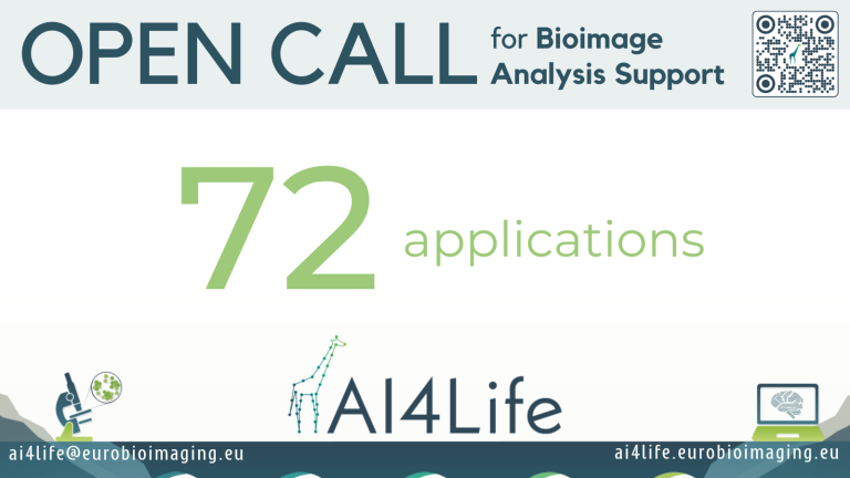
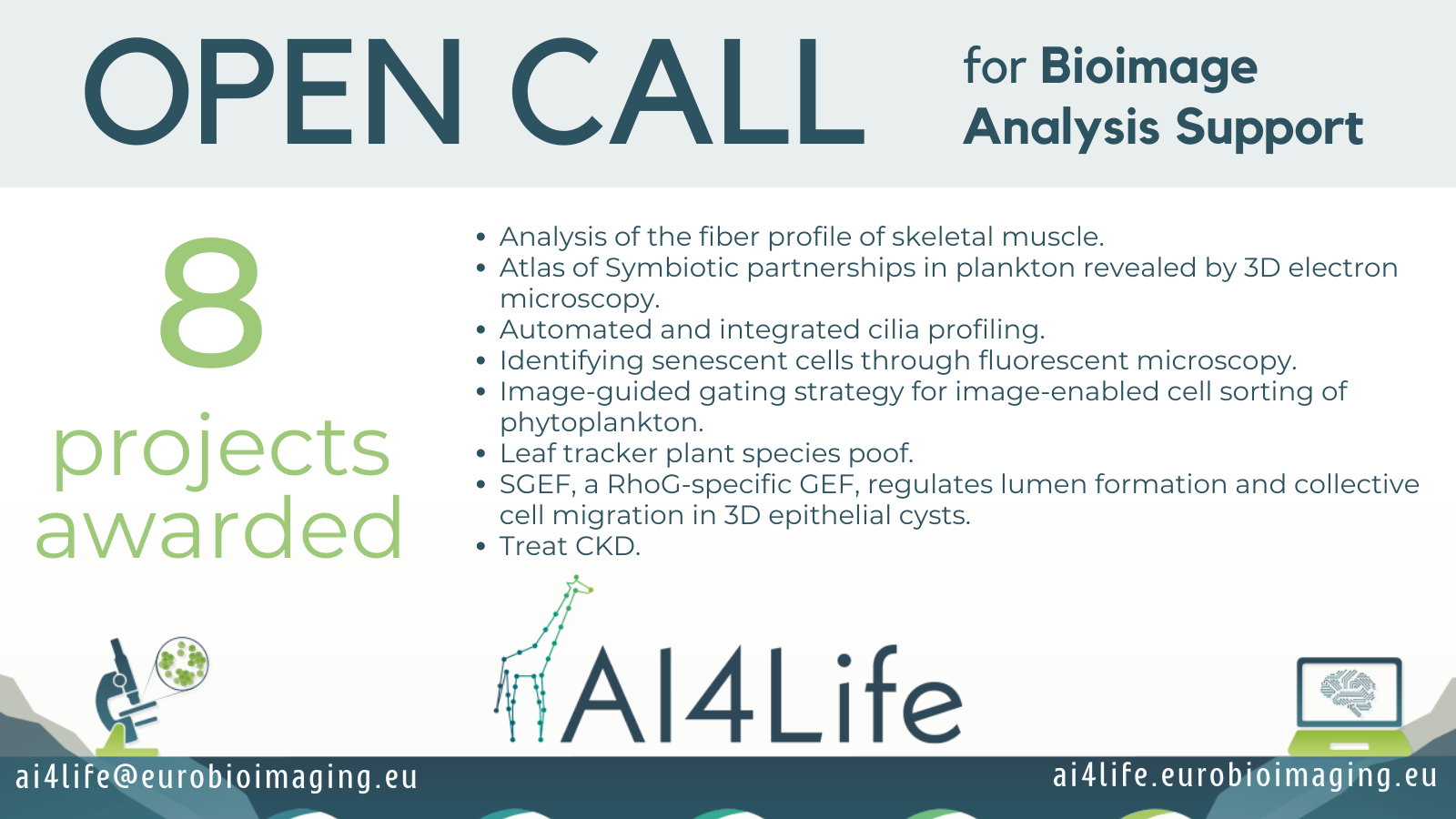
The first AI4Life Open Call received an impressive response, with a total of seventy-two applications. It proved to be an incredible opportunity for both life scientists seeking image analysis support and computational scientists eager to explore the evolving landscape of AI methodologies. In this blog post, we announce the awarded projects and invite you to join us behind the scenes as we explore the selection process that determined which projects have been selected.
First things first, here is the list of titles of the selected projects (in alphabetical order):
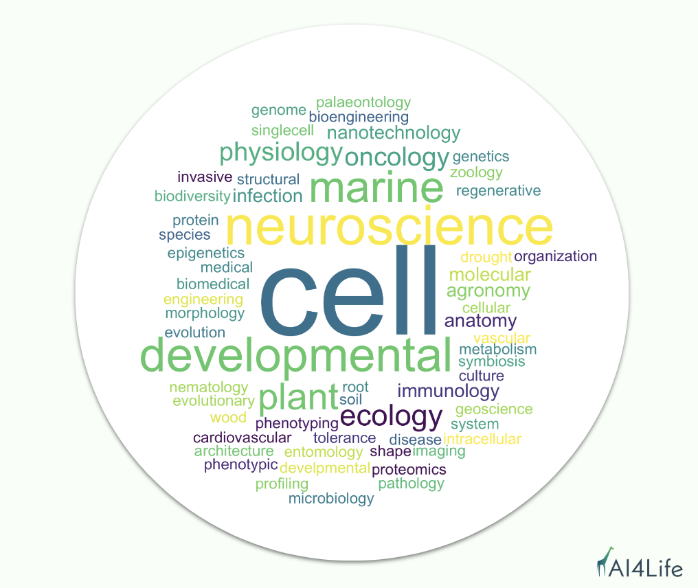
The projects are diverse, covering scientific topics ranging from Plant Biology, Physiology, Metabolism, Cell Biology, Molecular Biology, Marine Biology, Flow Cytometry, Medical Biology, Regenerative Biology, Neuroscience, etc. The researchers who have proposed the projects come from the following countries: France (2x), Germany, Italy, Netherlands, Portugal, and the USA (2x).
The selection procedure started with internal eligibility checks. Is the project submitted completely? Is the information complete and telling a complete story that is fit for external reviews? At this stage, we only had to drop 10 projects of a grant total of 72 submitted projects. Our intention was to only filter projects that drew an incomplete picture and leave the judgement of the scientific aspects to our reviewers.
After assembling a panel of 16 international reviewers (see list below), we distributed anonymized projects among them. All personal and institutional information was removed, only leaving project-relevant data to be reviewed. We aimed at receiving 3 independent reviews per project, requiring each review to review about 11 projects total.
Here is the list of questions we asked our reviewers via an electronic form:
Due to the unforeseen unavailability of some reviewers, we ended up with about 2.7 reviews per project, with some projects receiving 2 but most projects receiving all 3 desired reviews.
We first aggregated all reviews per project by averaging numerical values and concatenating textual evaluations. We then developed three project scores: a quality score (main metric), a total effort score, and a slightly more subjective excitingness score.
The final score was computed by: 0.75*(quality/effort) + 0.25*excitingness
This formula favors projects that are estimated to be conducted in less time, which is in line with our aim to help more individuals through the AI4Life Open Calls.
The selected projects will be assigned to our AI4Life experts waiting to support them. All other projects are offered a space in the AI4Life Bartering Corner, a new section soon to appear on our website, where projects will be showcased to computational experts who can reach out to the proposing parties and engage in a fruitful collaboration.
If you did not apply to the first Open Call, we invite you to do so at the beginning of 2024. Subscribe to our newsletter, we will inform you when the next call opens.
Additionally, if you are interested to put any open analysis problem you have on our Bartering Corner, please fill out this form.
If you need help quicker, we recommend Euro-BioImaging’s Web Portal, where you can access a network of experts in the field of image analysis. Please note that this service may involve associated costs, but access funds for certain research topics are available through initiatives such as ISIDORe.
by Caterina Fuster-Barceló
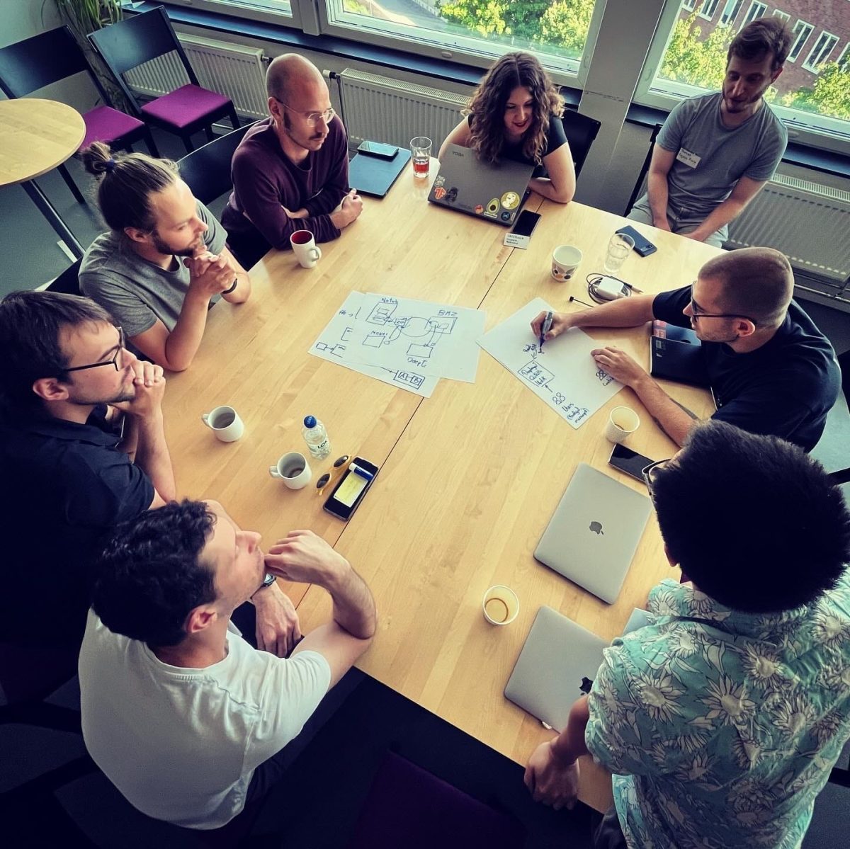
The AI4Life Hackathon on Web and Cloud Infrastructure for AI-Powered BioImage Analysis recently took place at SciLifeLab in Stockholm, Sweden. Organized by Wei Ouyang of KTH Sweden, in partnership with AI4Life and Global BioImaging, the event aimed to bring together experts in the field to discuss and design advanced web/cloud infrastructure for bioimage analysis using AI tools. Participants from both academia and industry worldwide attended, showcasing platforms like BioImage Model Zoo, Fiji, ITK, Apeer, Knime, ImJoy, Piximi, Icy, and deepImageJ. Read more in this article written by the project partners in FocalPlane.

by Caterina Fuster-Barceló
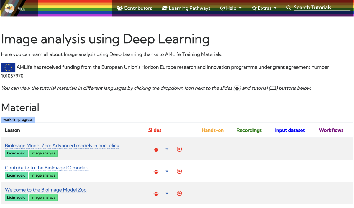
In an exciting collaboration, AI4Life has joined forces with the Galaxy Training Network (GTN) project to revolutionize the way researchers access training materials. The GTN, known for its dedication to promoting FAIR (Findable, Accessible, Interoperable, and Reusable) and Open Science practices globally, now incorporates AI4Life to expand its training offerings.
Through this collaboration, BioImage Model Zoo (BMZ) and AI4Life trainers have developed videos and slides to introduce the community to the BMZ, demonstrate proper utilization, and guide contributions. This exciting development allows the BMZ to reach a wider audience within the research community and offers a simplified, visual approach to understanding and utilizing the BMZ.
This collaboration between the BMZ and GTN opens up new opportunities for researchers to access training materials and gain a better understanding of the BMZ’s capabilities. By making the process more accessible and intuitive, the BMZ aims to facilitate its adoption among researchers from diverse backgrounds.
The integration of the BMZ into the GTN project represents a significant advancement in training resources, empowering researchers worldwide and fostering collaboration within the scientific community. Stay tuned for upcoming training materials that will unlock the full potential of the BMZ for your research pursuits.

by Estibaliz Gómez-de-Mariscal
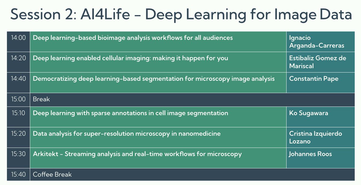
The 5th NEUBIAS Conference took place in Porto during the week of May 8th, 2023. It brought together experts in BioImage Analysis for the Defragmentation Training School and the Open Symposium. AI4Life actively participated in the event, contributing to both parts and covering topics from zero code Deep Learning tools, the Bioimage Model Zoo, BiaPy, Segment Anything for Microscopy, among others. Estibaliz Gómez-de-Mariscal has written a post in FocalPlane summarising the discussions and outcomes.

by Caterina Fuster-Barceló
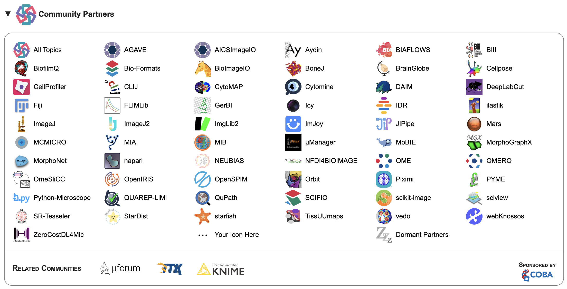
The BioImage Model Zoo (BMZ) has been incorporated as a Community Partner of the Image.sc forum, a discussion forum for scientific image software sponsored by the Center for Open Bioimage Analysis (COBA). The BMZ is a repository of pre-trained deep learning models for biological image analysis, and its integration into the Image.sc forum will provide a platform for the community to discuss and share knowledge on a wide range of topics related to image analysis.
The Image.sc forum aims to foster independent learning while embracing the diversity of the scientific imaging community. It provides a space for users to access a wide breadth of experts on various software related to image analysis, encourages open science and reproducible research, and facilitates discussions about elements of the software. All content on the forum is organized in non-hierarchical topics using tags, such as the “bioimageio” tag, making it easy for people interested in specific areas to find relevant discussions.
As a Community Partner, the BMZ joins other popular software tools such as CellProfiler, Fiji, ZeroCostDL4Mic, StarDist, ImJoy, and Cellpose, among others. The partnership means that the BMZ will use the Image.sc forum as a primary recommended discussion channel, and will appear in the top navigation bar with its logo and link.
The Image.sc forum has been cited in scientific publications, and users may reference it using the following citation:
Rueden, C.T., Ackerman, J., Arena, E.T., Eglinger, J., Cimini, B.A., Goodman, A., Carpenter, A.E. and Eliceiri, K.W. “Scientific Community Image Forum: A discussion forum for scientific image software.” PLoS biology 17, no. 6 (2019): e3000340. doi:10.1371/journal.pbio.3000340
The integration of the BMZ into the Image.sc forum will undoubtedly facilitate knowledge-sharing and collaborative efforts in the field of biological image analysis, benefiting researchers, developers, and users alike.

by Beatriz Serrano-Solano & Florian Jug
We are thrilled to announce that we received an impressive number of 72 applications to the first AI4Life Open Call!
The first AI4Life Open Call was launched in mid-February and closed on March 31st, 2023. It is part of the first of a series of three that will be launched over the course of the AI4Life project.
AI4Life involves partners with different areas of expertise, and it covers a range of topics, including marine biology, plant phenotyping, compound screening, and structural biology. Since the goal of AI4Life is to bridge the gap between life science and computational methods, we are delighted to see so much interest from different scientific fields seeking support to tackle scientific analysis problems with Deep Learning methods.
We noticed that the most prominent scientific field that applied was cell biology, but we also received applications from neuroscience, developmental biology, plant ecology, agronomy, and marine biology, as well as aspects of the medical and biomedical fields, such as cardiovascular and oncology.
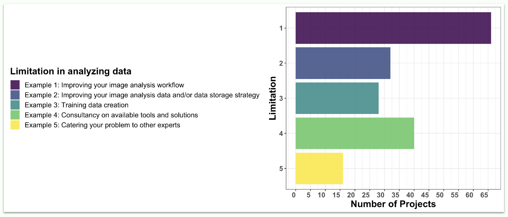
Applications were classified based on the type of problem that will need to be addressed. We found that improving the applicant’s image analysis workflow was the most common request (67 applications out of 72). This was followed by improving image analysis data and/or data storage strategy, training data creation, and consultancy on available tools and solutions. Of course, we also welcomed more specific challenges that were not falling within any of those categories.
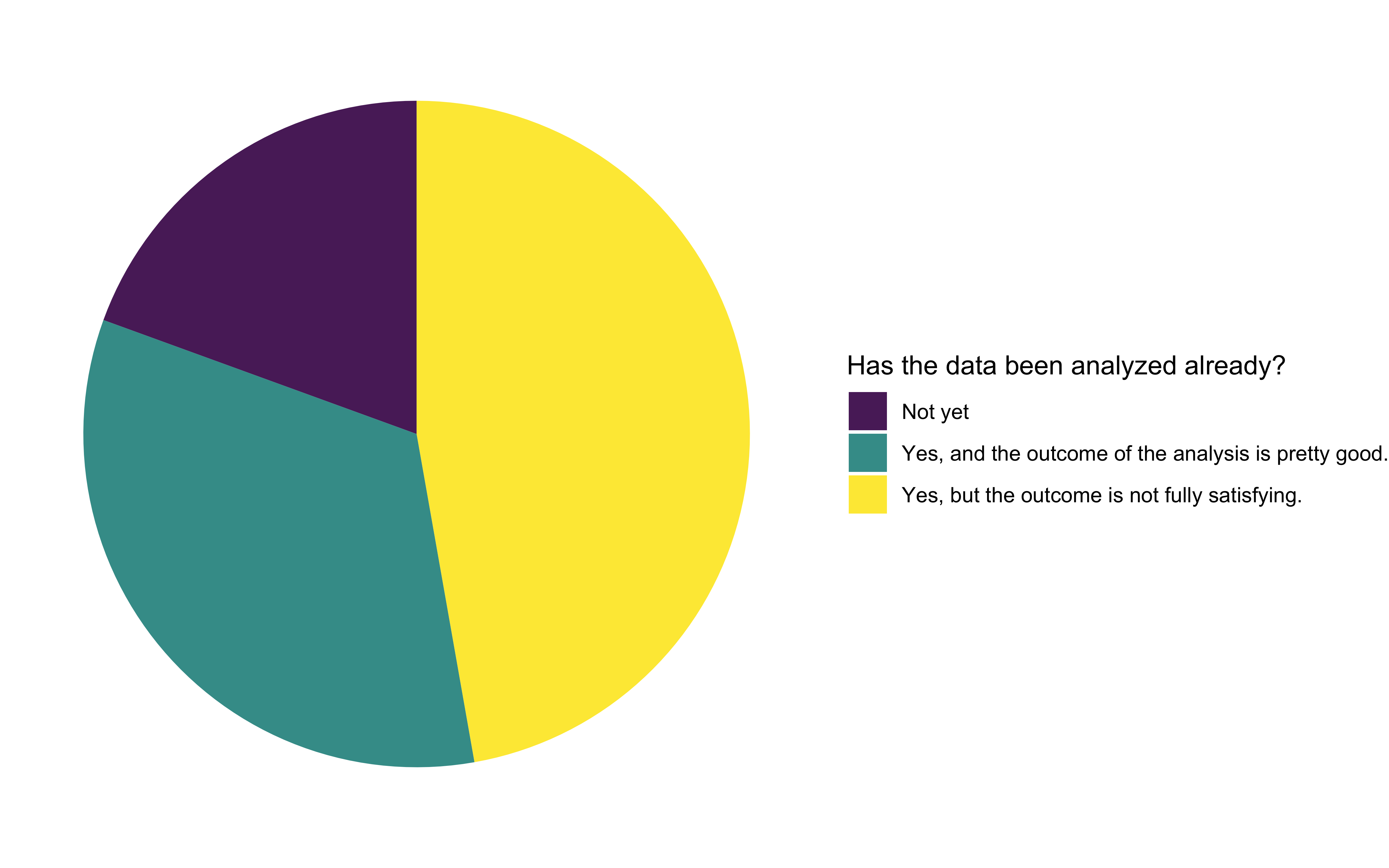
We were pleased to see that most of the applicants had already analyzed the data, but half of them were not fully satisfied with the outcomes. We interpreted this as an opportunity to improve existing workflows. The other half of the applicants were satisfied with their analysis results, but longed for better automation of their workflow, so it becomes less cumbersome and time-consuming. Around 20% of all applications haven’t yet started analyzing their data.
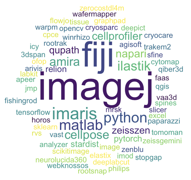
When asked about the tools applicants used to analyze their image data, Fiji and ImageJ were the most frequently used ones. Custom code in Python and Matlab is also popular. Other frequently used tools included Napari, Amira, Qupath, CellProfiler, ilastik, Imaris, Cellpose, and Zen.
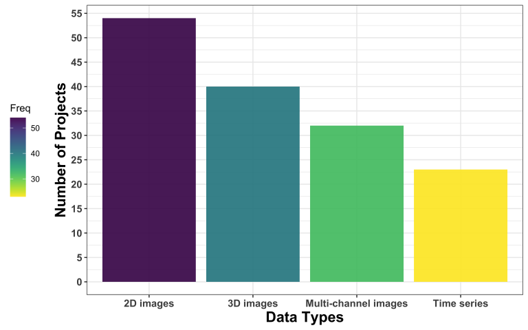
The most common kind of image data are 2D images, followed by 3D images, multi-channel images, and time series.
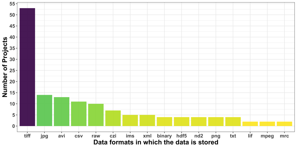
Regarding data formats, TIFF was the most popular one, followed by JPG, which is borderline alarming due to the lossy nature of this format. AVI was the third most common format users seem to be dealing with. CSV was, interestingly, the most common non-image data format.
Additionally, we asked about the relevance of the metadata to address the proposed project. 17 applicants didn’t reply, and 24 others did, but do not think metadata is quite relevant to the problem at hand. While this is likely true for the problem at hand, these responses show that the reusability and FAIRness of acquired and analyzed image data is not yet part of the default mindset of applicants to our Open Call.
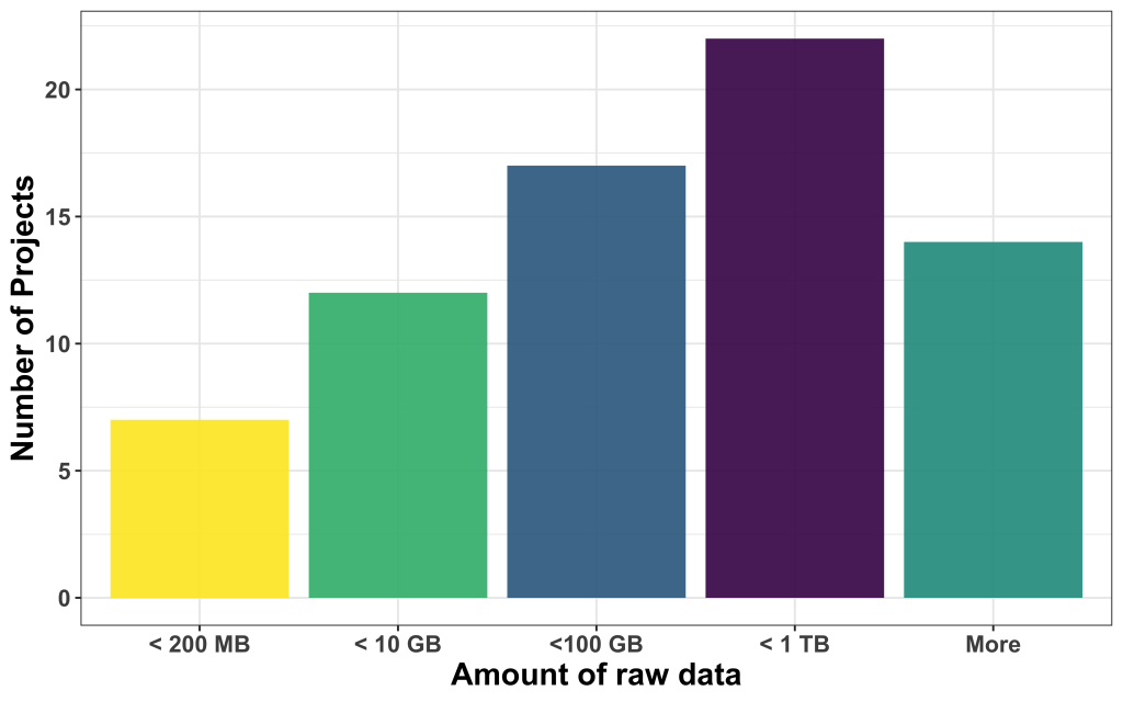
We found that most of the data available was large, with most of them in the range between 100 and 1000 GB, followed by projects that come with less than 10 GB, between 10 and 100 GB, and less than 200 MB. 14 projects had more data than 1 TB to offer.
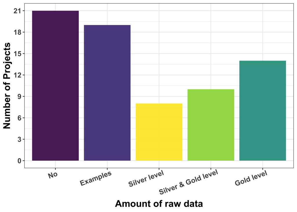
We also asked about the availability of labelled data and provided some guidelines regarding the kind and quality of such labels. We distinguished: (i) Silver ground truth, i.e. results/labels good enough to be used for publication (but maybe fully or partly machine-generated, and (ii) gold ground truth, i.e. human curated labels of high fidelity and quality.
The majority of applicants (40) had no labelled data or only very few examples. The rest have silver level (8), a mix of silver and gold levels (10) and gold level (14) ground truth available. The de-facto quality of available label data is, at this point in time, not easy to be assessed, but our experience is that users who believe to have gold-level label data are not always right.
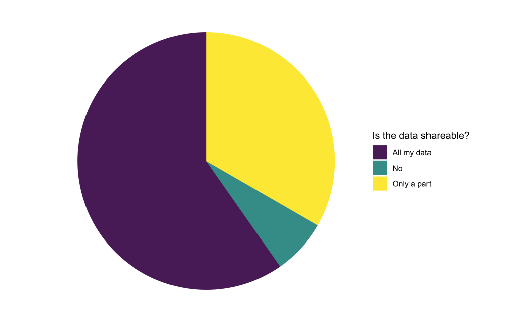
To train Deep Learning models, the computational experts will need access to the available image data. We found that only a small portion of applicants were not able to share their data at all. The rest is willing to share either all or at least some part of their data. When only parts of the data are sharable, reasons were often related to data privacy issues or concerns about sharing unpublished data.
We are currently undergoing an eligibility check and the pool of reviewers will start looking at the projects in more detail. In particular, they will rank the projects based on the following criteria:
The reviewers will also identify the parts of the project that can be improved, evaluate if deep learning can be of help and provide an estimation of the time needed to support the project.
We will keep you posted about all developments. Thanks for reading thus far! 🙂